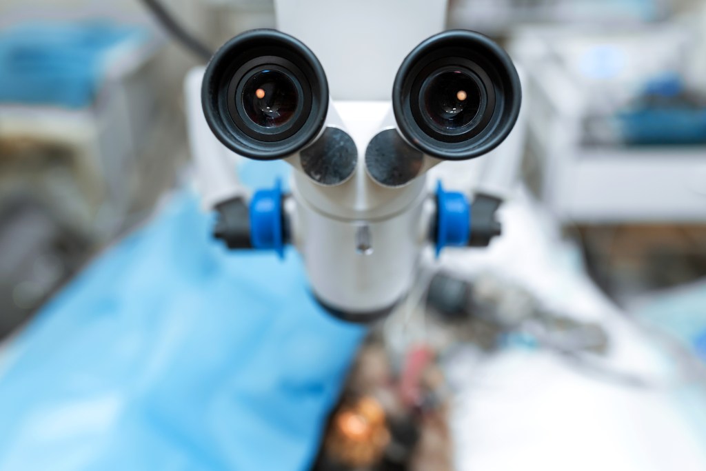
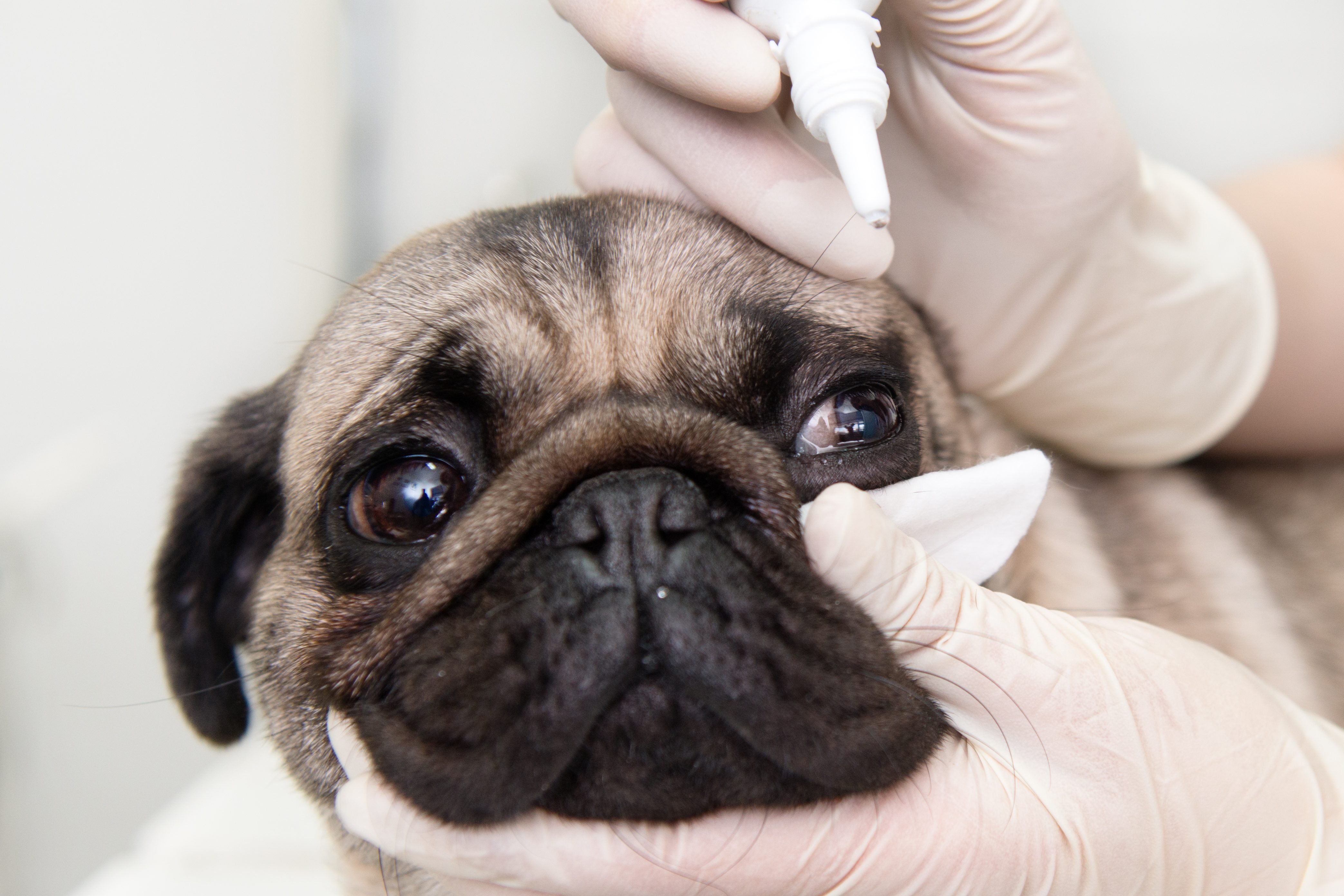
What is a board certified veterinary ophthalmologist?
A veterinary ophthalmologist specializes in diseases that may affect your pet’s eye and their vision. In order to become a board certified veterinary ophthalmologist, a veterinarian typically will complete an internship program followed by a residency, which results in a total of 8 to 10 years of post-college medical education. After completion of an ophthalmology residency, the candidate must then pass a rigorous 3-day, 5-part examination in order to become board certified by the American College of Veterinary Ophthalmologists. This process is very similar to the pathway that human medical doctors take to become board certified.
Some eye conditions can often be managed by your pet’s primary care veterinarian. However, many eye diseases require the attention of an ophthalmologist for advanced medical and surgical treatment.
What to expect during an ophthalmology consultation
The initial consultation typically lasts 20-30 minutes and includes measurement of tear production, assessment for corneal ulcers, evaluation of eye pressure, and a comprehensive examination of the entire eye. After the evaluation, the ophthalmologist will discuss your pet’s condition, treatment options, and, if needed, any additional recommended testing. After the consultation is completed, your primary care veterinarian will receive a referral document that discusses the diagnosis and treatment plan so that there is continuity of care.
If a non-emergent surgical procedure is needed, it will be scheduled for a later date, and, in most instances, your pet will go home the same day of surgery.


ADVANCED OPHTHALMIC DIAGNOSTIC TESTING:
This is not a complete list, and additional, less common procedures are also performed
- Eye certification examinations for breeding
- Exotic animal ophthalmic examinations
- Wildlife ophthalmology
- Slit-lamp biomicroscopy
- Funduscopy (retinal evaluation)
- Qualitative tear film assessment
- Schirmer tear test assessment
- Tonometry
- Gonioscopy
- Electroretinography
- Ocular ultrasound (including high frequency)
- Biopsy and ocular histopathology
- Dacryocystorhinography
- Computed tomography (CT scan)
- Magnetic Resonance Imaging (MRI scan)
ADVANCED NON-SEDATED PROCEDURES:
These procedures are often performed the same day without the need for any sedation or anesthesia
- Nasolacrimal irrigation
- Aqueocentesis
- Corneal and conjunctival biopsy
- Diamond burr debridement
- Corneal and conjunctival cytology
ADVANCED SURGICAL PROCEDURES:
- Phacoemulsification (cataract removal)
- Glaucoma surgery (endoscopic cyclophotocoagulation)
- Glaucoma surgery (artificial valve placement)
- Luxated lens removal (intracapsular lens extraction)
- Globe proptosis repair
- Entropion repair
- Eyelid reconstruction for severe injuries or tumors
- “Cherry eye” repair
- Cryosurgery for eyelid tumors (no general anesthesia)
- Keratectomy
- Conjunctival graft for eye rupture
- Perforated (ruptured) globe repair
- Corneoconjunctival transposition
- Corneal sequestrum surgery (corneoconjunctival transposition)
- Keratoleptynsis for corneal endothelial degeneration
- Corneal laceration repair
- Enucleation (eye removal)
- Intrascleral prosthesis placement in dogs
- Orbitotomy for tumor removal from behind the eye
- Parotid duct transposition
- Intraocular mass removal


